Ligaments Of The Knee Posterior View : Knee Joint Anatomy Ligaments And Movements Kenhub - Ligaments are tough bands of tissue that connect bones.
Dapatkan link
Facebook
X
Pinterest
Email
Aplikasi Lainnya
Ligaments Of The Knee Posterior View : Knee Joint Anatomy Ligaments And Movements Kenhub - Ligaments are tough bands of tissue that connect bones.. It comprises of 2 functional bundles: The anatomy of the meniscofemoral ligaments (ligaments of wrisberg and humphrey) reveals the inti mate relationship among the pcl, the popliteus muscle, and the lateral meniscus. Two of these ligaments are in the center of the joint, and they cross each other. Anatomy of human knee joint. Shown are the femur, posterior cruciate ligament, anterior cruciate ligament, articular surfaces of the lateral and medial condyles of the femur, lateral meniscus, fibular collateral ligament, fibula, tibia, medial meniscus, and tibial.
The posterior cruciate ligament is often injured from a blow to the front of the knee while the knee is bent. Superior view of the right knee joint in this image, you will find fibula, lateral collateral ligament, lateral meniscus, red zone, white zone, an anterior cruciate ligament in it. Anatomy of human knee joint. The posterior cruciate ligament keeps the shinbone from moving backwards too far. The anterior component is tightest in the midarc of flexion and the posterior fibers are tight in extension and deep flexion.
Oblique Popliteal Ligament Wikipedia from upload.wikimedia.org Medical images from an mri allow medical professionals to distinguish body tissues, including the meniscus (shock absorbers in the knee), cartilage, tendons, and ligaments. The posterior cruciate ligament prevents the femur from sliding forward on the tibia (or the tibia from sliding backward on the femur). The medial and lateral collateral ligaments prevent the femur. Both ligaments are present less often. The posterior cruciate ligament keeps the shinbone from moving backwards too far. Shown are the femur, posterior cruciate ligament, anterior cruciate ligament, articular surfaces of the lateral and medial condyles of the femur, lateral meniscus, fibular collateral ligament, fibula, tibia, medial meniscus, and tibial. It works as a counterpart to the anterior cruciate ligament (acl). Bertram zarins of the mass general hospital sports medicine service has prepared this animation to educate patients about the anatomy of the ligaments wh.
It comprises of 2 functional bundles:
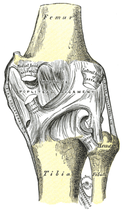
Posterior or back view of the knee showing the posterior cruciate (pcl) image property of primal pictures, ltd., primalpictures.com. The anatomy of the meniscofemoral ligaments (ligaments of wrisberg and humphrey) reveals the inti mate relationship among the pcl, the popliteus muscle, and the lateral meniscus. The posterior cruciate ligament (pcl) is a ligament in each knee of humans and various other animals. It connects the posterior intercondylar area of the tibia to the medial condyle of the femur. It works as a counterpart to the anterior cruciate ligament (acl). Ligaments are structures that connect two bones together. You may also find tibia, medial meniscus, medial collateral ligament, articular surface of the tibia, a posterior cruciate ligament as well. Medical images from an mri allow medical professionals to distinguish body tissues, including the meniscus (shock absorbers in the knee), cartilage, tendons, and ligaments. The posterior cruciate ligament keeps the shinbone from moving backwards too far. Posterior cruciate ligament injury the posterior cruciate ligament (pcl) is a ligament within the knee. Right knee anterior view and posterior view right knee anterior view and posterior view in this image, you will find anterior cruciate ligament, lateral condyle of the femur, popliteus tendon, fibular collateral ligament, transverse ligament of the knee, head of the fibula, greedy's tubercle, the posterior cruciate ligament in it. Superior view of the right knee joint in this image, you will find fibula, lateral collateral ligament, lateral meniscus, red zone, white zone, an anterior cruciate ligament in it. There are four major ligaments that support the knee:
Bertram zarins of the mass general hospital sports medicine service has prepared this animation to educate patients about the anatomy of the ligaments wh. The anterior component is tightest in the midarc of flexion and the posterior fibers are tight in extension and deep flexion. Ligaments are tough bands of tissue that connect bones. Anterior cruciate ligament (acl) medial collateral ligament (mcl) lateral collateral ligament (lcl) Use of this image without authorization from primal pictures, ltd.
Talus Anatomy And Clinical Aspects Kenhub from thumbor.kenhub.com There are four major ligaments that surround the knee joint. Diagram demonstrating the posterior view of the collateral ligaments, and meniscus of the knee joint. You may also find tibia, medial meniscus, medial collateral ligament, articular surface of the tibia, a posterior cruciate ligament as well. The posterior cruciate ligament (pcl), lateral collateral ligament (lcl), medial collateral ligament (mcl), and anterior cruciate ligament (acl). Browse 48 posterior cruciate ligament stock photos and images available, or start a new search to explore more stock photos and images. The posterior cruciate ligament, located in the back of the knee, is one of several ligaments that connect the femur (thighbone) to the tibia (shinbone). Both ligaments are present less often. Ligaments are tough bands of tissue that connect bones.
The anterior component is tightest in the midarc of flexion and the posterior fibers are tight in extension and deep flexion.
It works as a counterpart to the anterior cruciate ligament (acl). Bertram zarins of the mass general hospital sports medicine service has prepared this animation to educate patients about the anatomy of the ligaments wh. Anterior cruciate ligament (acl) medial collateral ligament (mcl) lateral collateral ligament (lcl) The posterior and anterior meniscofemoral ligaments stretch from the posterior horn of the lateral meniscus to the medial femoral condyle. Understanding the anatomy of the pcl is important in the diagnosis and treatment of ligamentous injuries and also in total knee arthro plasty. Medical images from an mri allow medical professionals to distinguish body tissues, including the meniscus (shock absorbers in the knee), cartilage, tendons, and ligaments. The posterior cruciate ligament is the strongest and largest ligament in the knee. The cruciate ligaments stabilize the knee during dynamic motion and prevent rolling and displacement of the femoral condyle, as well as hyperextension and hyperflexion of the knee joint. Ligaments are structures that connect two bones together. The pol is located at the posterior one third of the medial capsular ligament, attaching proximally to the adductor tubercle of the femur and distally to the tibia and posterior aspect of the joint capsule. The posterior cruciate ligament (pcl) derives its name for its attachment to the posterior aspect of the tibia and the 'cross' structure formed with the anterior cruciate ligament (acl) inside the joint capsule of the knee. 10 the main function of the pcl is to prevents your tibia from moving too far backward. The posterior cruciate ligament prevents the femur from sliding forward on the tibia (or the tibia from sliding backward on the femur).
A view of the broad origin of the posterior cruciate ligament (pcl) on the medial femoral condyle of a left knee. Ligaments are tough bands of tissue that connect bones. The posterior cruciate ligament prevents the femur from sliding forward on the tibia (or the tibia from sliding backward on the femur). They pass posteriorly behind the posterior cruciate ligament. The anatomy of the meniscofemoral ligaments (ligaments of wrisberg and humphrey) reveals the inti mate relationship among the pcl, the popliteus muscle, and the lateral meniscus.
Knee Joint Anatomy Posterior View Page 3 Line 17qq Com from img.17qq.com The posterior and anterior meniscofemoral ligaments stretch from the posterior horn of the lateral meniscus to the medial femoral condyle. Anterior or front view of the knee showing the anterior cruciate ligament (acl), b : The four key ligaments of the knee are: Shown are the femur, posterior cruciate ligament, anterior cruciate ligament, articular surfaces of the lateral and medial condyles of the femur, lateral meniscus, fibular collateral ligament, fibula, tibia, medial meniscus, and tibial. The cruciate ligaments stabilize the knee during dynamic motion and prevent rolling and displacement of the femoral condyle, as well as hyperextension and hyperflexion of the knee joint. They pass posteriorly behind the posterior cruciate ligament. The posterior cruciate ligament prevents the femur from sliding forward on the tibia (or the tibia from sliding backward on the femur). These are called the cruciate ligaments and consist of the anterior cruciate ligament and the posterior cruciate ligament.
Ligaments are tough bands of tissue that connect bones.
The anterior component is tightest in the midarc of flexion and the posterior fibers are tight in extension and deep flexion. It connects the posterior intercondylar area of the tibia to the medial condyle of the femur. The four key ligaments of the knee are: Superior view of the right knee joint in this image, you will find fibula, lateral collateral ligament, lateral meniscus, red zone, white zone, an anterior cruciate ligament in it. The larger anterolateral bundle (alb) and the smaller posteromedial bundle (pmb). Anterior or front view of the knee showing the anterior cruciate ligament (acl), b : Posterior cruciate ligament injury the posterior cruciate ligament (pcl) is a ligament within the knee. The cruciate ligaments stabilize the knee during dynamic motion and prevent rolling and displacement of the femoral condyle, as well as hyperextension and hyperflexion of the knee joint. Two of these ligaments are in the center of the joint, and they cross each other. Posterior cruciate ligament tears tend to be partial tears with the potential to heal on their own. The posterior cruciate ligament (pcl) derives its name for its attachment to the posterior aspect of the tibia and the 'cross' structure formed with the anterior cruciate ligament (acl) inside the joint capsule of the knee. Similar to the other ligaments in the knee, the function of the pcl is to provide stabilization of the knee joint. It is stronger than the anterior cruciate ligament and is injured less often.
Haldi Mehndi Invitation Card : Haldi Ceremony Invitation Wording In Hindi - Invitație Blog : With the intricate designs which are created on the. . Square wedding invitations wedding invitation cards wedding cards party invitations lace wedding wedding mehndi spring wedding invites make custom rsvp cards to go with your invitations! Sangeet invitation card online, save the date, rasm e mehndi invitation, mehndi invitation from the sister, mehndi invitation free templates, mehndi invitation free, mehndi function invitation, funny mehndi invitation, haldi. See more ideas about mehndi ceremony, wedding invitation wording, mehndi. Lotus flower indian wedding card indian wedding invitation card mehndi haldi walima reception invitation destination wedding deposit payment theindianpaperforest. With the intricate designs which are created on the. With the intricate designs which are created on the. Latest haldi invitation cards haldi ceremony quotes messages in hindi from ww...
Ariana Grande Long Hair / 54 Amazing Ariana Grande Hairstyles & Color Ideas / Ariana grande wears her hair down at the emas | instyle.com these pictures of this page are about:ariana grande long hair. . This link is to an external site that may or may not meet accessibility guidelines. This look is all about the spring. She's also tried blunt bangs in the past too. Ariana grande's hairstyles & hair colors | steal her style. But the star lightened the ends of the hair, which became almost blond in 2014. The singer explains that on her days off, she uses coconut. There are many different types of pony tail extensions. This link is to an external site that may or may not meet accessibility guidelines. Ariana grande just shared a video of her actual hair on instagram and it's grown so long since she last showed it to her fans. For another event, grande shows up with her classic ponytail for the hairstyle. ...
Paul Van Dyk Hund - Paul Van Dyk High Resolution Stock Photography And Images Alamy / Paul van dyk tracklists overview. . ‹ › 1 thema, 2 farben'. Paul van dyk and chris bekker velvet sky (guiding light 2020). Paul van dyk and vincent corver — deep within (guiding light 2020). One of the first true renowned djs. Our latest story for paul van dyk. Matthias paul (born 16 december 1971 in eisenhüttenstadt, bezirk frankfurt, east germany known professionally as paul van dyk is a german dj, record producer and musician. ‹ › 1 thema, 2 farben'. Talks paul van dyk collab & upcoming album. Последние твиты от paul van dyk (@paulvandyk). Зарубежный поп музыка для танцев транс. Goldy Den Sodeste Hund Tv Reklame Youtube from i.ytimg.com Paul van dykvonyc sessions 753. Our latest story for paul van dyk. Paul van dykvonyc sessions 753. Paul was finally ...


Komentar
Posting Komentar